

Pregnancy is an exciting time, filled with many questions and new experiences. Among these is the process of undergoing a pregnancy ultrasound. These routine procedures provide valuable information about both the mother and the baby. Non-invasive imaging technology is widely used in prenatal care. It has become an integral tool in monitoring pregnancies worldwide.
What Are Pregnancy Ultrasounds?
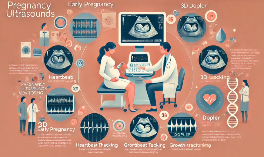
Pregnancy ultrasounds use high-frequency sound waves to create images of the baby and uterus. This technology is non-invasive and poses no known risks to mothers or babies. During the scan, a device called a transducer is placed on the abdomen or inserted into the vaginal canal, depending on the stage of the pregnancy. The sound waves bounce off tissues and fluids, creating a detailed image on a screen.
These images allow healthcare providers to gather information about the pregnancy. Ultrasounds can show the baby’s position, the amount of amniotic fluid, and even the placenta’s location. Depending on the stage of the pregnancy, they may also reveal specific details about the baby’s growth and development.
Why Are They Needed?
A pregnancy ultrasound plays an integral role in prenatal care for a variety of reasons. Ultrasounds allow healthcare providers to monitor fetal development and detect issues early. This can include identifying potential complications related to fetal growth, placenta position, or the baby’s overall health. Identifying these details early can guide healthcare providers in tailoring prenatal care appropriately.
Ultrasounds can also provide a window into the baby’s environment. The scans allow doctors to assess the level of amniotic fluid and check for the presence of multiple pregnancies. Ultrasounds are a useful way to establish or confirm how far along the pregnancy is. This helps in predicting due dates or assessing whether the baby is growing as expected for its gestational age.
Pregnancy ultrasounds also offer reassurance to expectant mothers. Seeing the baby on the screen and receiving detailed information about the pregnancy can help reduce concerns. In some cases, additional imaging options may be recommended to ensure the baby’s development is progressing as expected.
When Should I Get a Pregnancy Ultrasound?
The timing of ultrasounds varies depending on the needs of the pregnancy. Healthcare providers typically schedule the first ultrasound early in pregnancy. This is often done between six and eight weeks to confirm the pregnancy and estimate the due date. The scan can detect the baby’s heartbeat and confirm whether it’s a single or multiple pregnancy.
A second ultrasound is usually performed around 18 to 22 weeks in what is sometimes called the anatomy scan. This detailed exam checks the baby’s growth and development, while evaluating the placenta and other factors. The anatomy scan typically includes a thorough look at the baby’s heart, brain, and other organs.
Some women may require additional ultrasounds later in pregnancy. These may focus on specific concerns, such as the baby’s position or weight. Common reasons for later ultrasounds can include checking for low amniotic fluid levels or cases of high-risk pregnancies.
Learn More About Your Prenatal Journey
Pregnancy ultrasounds are a valuable part of the prenatal experience for expectant mothers. They provide detailed insights into the baby’s growth and the health of the pregnancy, all in a safe and non-invasive way. If you have questions about pregnancy ultrasounds or your overall care, your healthcare provider is the best source of information. Follow their recommendations and ask about the specific scans that may benefit you and your baby.
- 4 Reasons To Schedule Regular Water Heater Maintenance
- Crawl Space Repairs: Local Case Studies from Belleville
- The Benefits of Primary Care for Preventing Future Health Issues
- The Impactful Addition of Higher Education for Your Resume: Elevate Career Prospects
- Am I a Good Candidate for Stem Cell Therapy?


